

Make Dental Work Easier
Bring the best Chinese professional dental equipment to Africa.
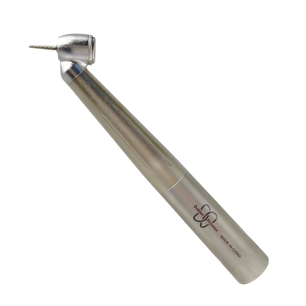
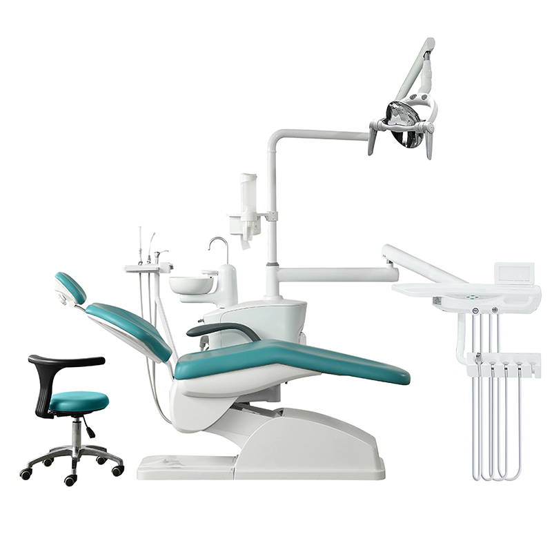
Golden Promise Dental:Brings the best Chinese dental products to Africa!

With advancements in technology shaping every facet of modern life, the field of dentistry is no exception. Digital operating microscopes have revolutionized dental practice, offering unmatched precision, effectiveness, and patient satisfaction. Curious to learn more about how these cutting-edge tools are transforming the world of dentistry? Read on to discover the benefits and innovations brought by digital operating microscopes in dentistry.
Enhanced Visualization and Precision
One of the most compelling advantages of digital operating microscopes in dentistry is the enhanced visualization they provide. Traditional dental procedures often rely heavily on the dentist's naked eye or basic magnification tools, which can sometimes pose limitations in visibility. In contrast, digital operating microscopes offer magnification levels ranging from 5x to 40x, allowing dentists to observe minute details that would otherwise go unnoticed.
The high-resolution imaging capabilities of these microscopes enable professionals to conduct more precise evaluations and procedures. For instance, intricate procedures such as root canal therapy or micro-surgeries can be executed with greater accuracy. The microscopes capture and display real-time images on a monitor, giving dentists a broader field of view and additional clarity, which directly contributes to better patient outcomes.
This enhanced visualization also aids in diagnosing conditions that might be missed with less advanced tools. Small fractures, caries, and periodontal issues can be detected early, facilitating timely intervention and preventing more severe complications down the line. As a result, patient diagnosis becomes more straightforward, exact, and reliable.
Additionally, these microscopes often feature adjustable lighting, providing optimal illumination during procedures. This minimizes shadows and inconsistencies in lighting, further enhancing the dentist's ability to see and work on the tiny, intricate spaces within the oral cavity. When paired with high-resolution imaging, this creates a potent combination that significantly increases the success rates of dental treatments.
Ergonomics and Reduced Strain
Dentistry can be physically demanding, often requiring practitioners to assume awkward positions for long periods. This can result in musculoskeletal strain and discomfort, which can inevitably affect the quality of dental care. Digital operating microscopes help mitigate these challenges by enabling dentists to work in more ergonomic positions.
Most digital operating microscopes come with adjustable features that allow the dentist to customize the height, angle, and position of the microscope according to their comfort. This reduces the need for constant bending and awkward contortions, promoting a healthier working posture. Moreover, the ability to view the operating field on a large monitor instead of through a small lens reduces eye strain and fatigue, making it easier for professionals to focus on the task at hand.
The ergonomic design of these advanced microscopes also translates to longer, more efficient work periods. Dentists can perform complex procedures without the interruption caused by physical discomfort, leading to increased productivity and patient turnover. This benefit extends beyond the individual practitioner to improve the overall efficiency and effectiveness of the dental practice.
By minimizing the physical toll on dentists, these tools also play a role in career longevity. Reducing the risk of repetitive strain injuries and other work-related conditions allows dentists to maintain a high level of performance over the years, ensuring that patients continue to receive quality care from experienced practitioners.
Patient Experience and Education
The use of digital operating microscopes in dentistry significantly enhances the patient experience. With the ability to display real-time images and video, patients are given a unique view of their dental condition, fostering better understanding and engagement. This method of visual communication helps demystify procedures, making patients feel more informed and at ease.
When patients can see the exact areas needing treatment and understand the rationale behind specific procedures, they are more likely to cooperate and adhere to treatment plans. This active involvement not only builds trust but also encourages better oral health habits long-term. For instance, seeing the extent of plaque buildup or gum disease firsthand can motivate patients to take better care of their teeth and gums.
Moreover, the high-quality imaging from digital operating microscopes aids in documenting and tracking progress over time. Dentists can take before-and-after images to show the effectiveness of treatments, which can be immensely satisfying and reassuring for patients. This visual documentation also facilitates better communication with dental insurers, making it easier to justify the necessity of treatments and secure coverage.
In addition to enhancing patient care during visits, digital operating microscopes serve as powerful educational tools for dental students and less experienced practitioners. The real-time imagery can be shared with an audience, enabling trainees to gain a clearer understanding of various procedures and techniques, thereby improving their educational experience.
Integration with Digital Dental Workflow
In the contemporary dental practice, seamless integration with digital workflows is essential. Digital operating microscopes are designed to be compatible with other dental technologies, such as CAD/CAM systems, digital X-rays, and patient management software. This integration ensures a more efficient, streamlined process from diagnosis to treatment and follow-up.
By combining the high-resolution imagery from digital microscopes with other digital diagnostic tools, dentists can create a comprehensive and highly accurate treatment plan. For instance, capturing detailed images of a patient's dental structure can assist in designing precise dental restorations using CAD/CAM technology. This close collaboration between diagnostic and restorative tools optimizes the accuracy and fit of prosthetics and other dental devices, ultimately improving patient outcomes.
Furthermore, the ability to store and share detailed images and videos enhances communication within the dental team. Complex cases can be more easily consulted with specialists, even remotely, as digital images and videos can be transmitted for review, facilitating more comprehensive and informed treatment planning.
The integration of digital operating microscopes in a practice also supports better record-keeping and documentation, ensuring that every step of the treatment process is recorded with precision. This not only complies with regulatory requirements but also serves as a valuable resource for future reference, quality control, and training purposes.
Innovations and Future Potential
The landscape of dental technology is continuously evolving, and digital operating microscopes are at the forefront of these advancements. Recent innovations have introduced features such as autofocus, image stabilization, and enhanced 3D visualization, taking dental practice to new heights of precision and effectiveness. These features not only simplify the use of microscopes but also offer enhanced diagnostic capabilities.
Autofocus technology, for instance, allows dentists to focus on specific areas quickly and accurately, reducing the time spent adjusting the microscope manually. Image stabilization helps maintain clear and steady visuals even during longer procedures, ensuring continuous high-quality imaging. These features allow for a more fluid and efficient workflow, further benefiting both dentists and patients.
The potential for artificial intelligence (AI) integration also holds promise for the future of digital operating microscopes in dentistry. AI algorithms could assist in real-time diagnostics, identifying potential issues and suggesting treatment options based on vast datasets. This would act as an invaluable second opinion, supplementing the dentist's expertise with data-driven insights.
Moreover, advancements in tele-dentistry could see the role of digital operating microscopes expand even further. With high-quality imaging, dentists could conduct remote consultations more effectively, diagnosing and planning treatment for patients in remote or underserved areas. This could significantly broaden access to specialized dental care, addressing disparities in healthcare availability.
Overall, the future of digital operating microscopes in dentistry looks exceedingly bright, with ongoing innovations and integrations poised to offer even greater benefits and possibilities.
In summary, digital operating microscopes have indeed revolutionized the field of dentistry by enhancing visualization, improving ergonomics, elevating patient experience and education, integrating smoothly with digital workflows, and driving innovations. These high-tech tools not only improve the quality of dental care but also make the process more efficient and comfortable for both dentists and patients alike.
As technology continues to evolve, we can anticipate even more groundbreaking advancements in this field, promising an exciting future for digital operating microscopes in dentistry. Adopting and mastering these innovative tools is not merely an option for modern dental practitioners but a necessity in delivering the best possible care.
Copyright © 2024 Golden Promise Dental Co.,Ltd. | All Rights Reserved
We are here to help you! If you close the chatbox, you will automatically receive a response from us via email. Please be sure to leave your contact details so that we can better assist

